
FORT WORTH, TX / ACCESS Newswire / November 19, 2026 / Vadzo Imaging, a provider of embedded vision camera products for OEMs and system integrators, today positions the Falcon-521MRS as a 5MP Digital Pathology Camera for monochrome tissue imaging and microscopic examination. Built on the Onsemi AR0521 sensor and part of Vadzo's USB camera portfolio, it combines monochrome output, 5MP high resolution, near-infrared response, a rolling shutter, and a USB 3.2 Gen 1 Type-C interface. Vadzo presents a 5MP Digital Pathology USB Camera for instrument makers, with guidance on sampling, illumination, and color reconstruction.
Why a 5MP Digital Pathology Camera Starts with Sampling and Light
Digital pathology replaces the eyepiece with a sensor. A tissue section a few microns thick, stained to show its structures, is imaged through a microscope objective, and the picture must keep the fine detail that a pathologist or an algorithm needs, such as the edge of a nucleus or the texture of a cell. Two things decide whether the detail survives. The first is sampling, which is how many pixels cover each feature. The second is light, which is how much of it each pixel collects and how evenly it is delivered across the field.
A color sensor loses some of both. Its color filter array lets each pixel see only one color, and software interpolates the rest, which softens fine edges, and the filters absorb a share of the light. A monochrome sensor avoids both losses. Pathology also needs more than brightfield color. Fluorescence work, unstained samples, and some research methods use near-infrared or single-channel images, and a scanner that handles many slides needs a camera that connects simply, uses standard drivers, and delivers consistent frames.
How a 5MP Digital Pathology Camera Resolves Tissue Detail at the Microscope
The Falcon-521MRS is built on the Onsemi AR0521, a 5MP (2592 x 1944) monochrome rolling shutter sensor with a 1/2.5 inch optical format and 2.2-micron pixels. A short calculation shows how the sensor meets the microscope. With a 20-times objective and a 1-times relay, each 2.2-micron pixel covers about 0.11 microns at the specimen, and the field measures about 285 by 214 microns. An objective with a numerical aperture of 0.75 resolves about 0.45 microns in green light, so the sensor samples the image more than four times finer than the optics require, and a lower magnification relay or a larger field can make better use of the pixels. A tissue area of 15 by 15 millimeters would need about 3,700 such fields, which shows why scanners tile the slide and why transfer speed matters.
Color is rebuilt by illumination. A monochrome camera can take three exposures of the same still field under red, green, and blue light, and software combines them into a color image. This sequential method gives full resolution in every channel, which a color sensor with a Bayer filter cannot, and it suits slides because the specimen does not move between exposures. It does require a stage that settles quickly, an illumination source that switches precisely, and a calibration of the color balance, and the device maker remains responsible for the accuracy of the color. Because the sensor reads the image row by row, the camera uses a rolling shutter, and still slides show no visible effect.
The camera connects over USB 3.2 Gen 1 with a Type-C connector, a nominal 5 gigabits per second. A 5MP frame at 8 bits per pixel is about 40 million bits, so 30 frames per second needs about 1.2 gigabits per second, and at 12 bits about 1.8, both comfortably inside the link, though real throughput is lower than the nominal rate. Near-infrared response supports fluorescence and some label-free methods, and teams should confirm the wavelength range of the camera against the dyes and filters they use. Teams should also confirm the output format, the exposure range, and the frame rates against the product documentation. A short test with a real stained slide, imaged under each color and compared with a reference image, shows how well the chain reproduces what the pathologist expects.
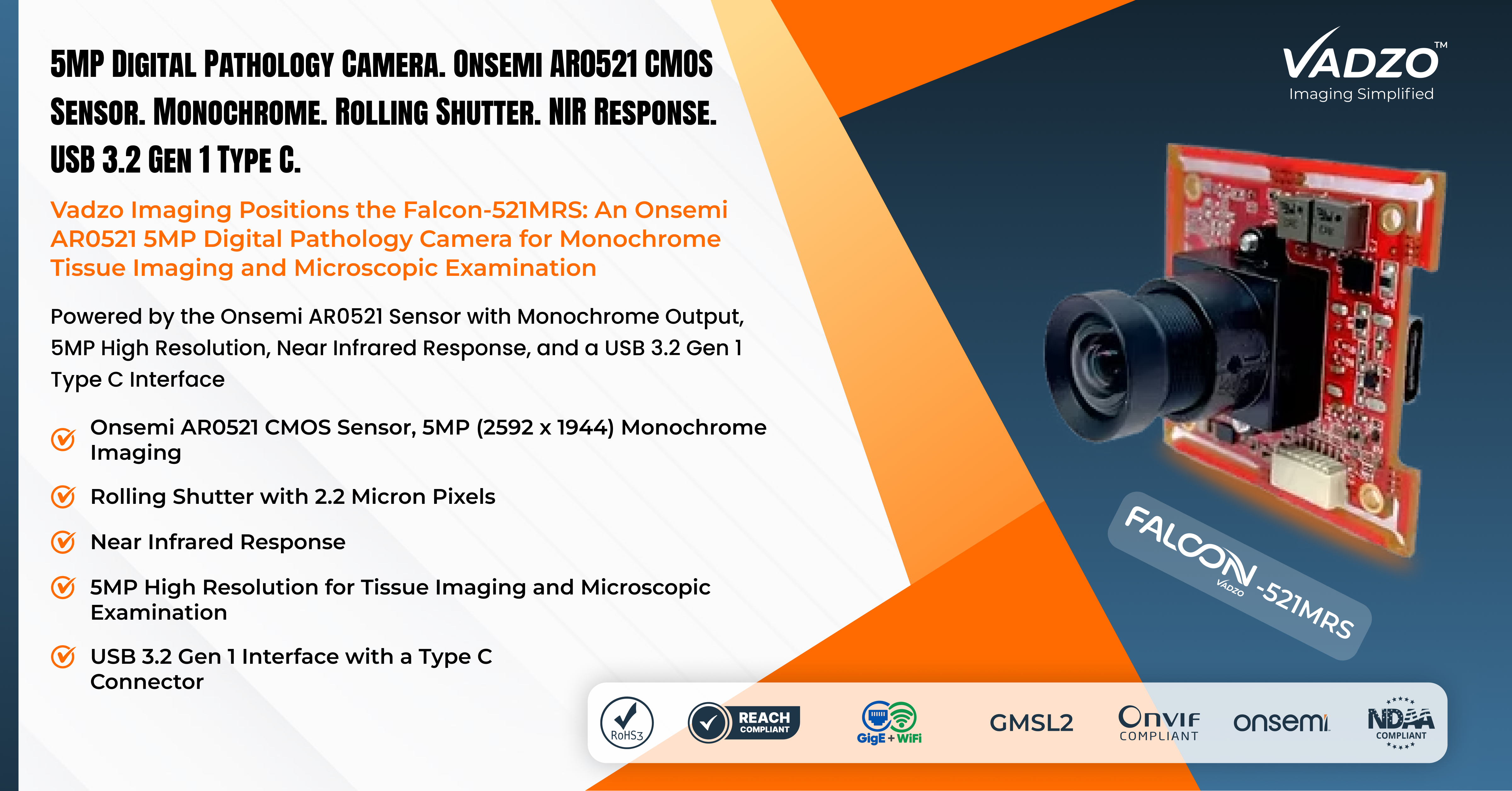
Product Overview: 5MP Digital Pathology Camera for Monochrome Tissue Imaging
The Falcon-521MRS is a 5MP monochrome USB module built on the Onsemi AR0521 sensor and positioned for digital pathology. As an AR0521 Monochrome Pathology USB Camera, it records brightness at every pixel, and as a 5MP NIR Pathology USB Camera, it responds in the near infrared. The 5MP Rolling Shutter USB Camera designation covers its readout, and the AR0521 Digital Pathology USB Camera designation covers its use behind a microscope. The module also serves as a 5MP Monochrome Pathology USB Camera, an AR0521 NIR USB Camera, a 5MP NIR USB Camera, a 5MP Mono Pathology USB Camera, a 5MP Tissue Imaging USB Camera, an AR0521 Pathology USB Camera, a 5MP Pathology Imaging USB Camera, a 5MP Monochrome Microscopy USB Camera, and an AR0521 Rolling Shutter Pathology USB Camera for designs that need a standard interface.
Key Capabilities of the 5MP Digital Pathology Camera
Key specs: 5MP (2592 x 1944) Max Resolution | Onsemi AR0521 1/2.5 inch Sensor Format | 2.2 micron Pixel | Monochrome with Rolling Shutter | USB 3.2 Gen 1 Type C Interface | Optics per Module Configuration
Monochrome Sensor Output
A monochrome sensor has no color filter array, so more light reaches each pixel, and no interpolation softens fine edges. For tissue imaging, this gives sharper structures than a color sensor of the same resolution, and a color image can be built by sequential illumination. Teams that need single-exposure color should use a color module from the same family.
Rolling Shutter Readout
The sensor reads the image row by row, which gives good resolution and sensitivity at reasonable cost. A slide on a settled stage holds still, so the effect is invisible. A stage that is still moving during the exposure can show skew, so teams should wait for the stage to settle or use a short exposure.
Near Infrared Response
The sensor responds in the near infrared, which supports fluorescence and some label-free methods, and it lets a system use wavelengths that penetrate tissue and avoid some interference. Sensitivity falls toward the longer wavelengths of silicon sensors, so teams should confirm the response at the wavelengths they use and match the filters to it.
5MP High Resolution
The 5MP frame of 2592 x 1944 pixels, with 2.2-micron pixels, puts plenty of pixels on tissue structures and covers a usable field. The module works as a 5MP USB Camera Module and a 5MP High Resolution USB Camera Module that joins Vadzo's USB range. As a 5MP Pathology USB Camera Supplier, Vadzo supports adapter matching, firmware customization, and enclosure design.
USB 3.2 Gen 1 Type C Interface
The module connects over USB 3.2 Gen 1 with a Type C connector and appears as a UVC device on Windows, Linux, and macOS without a proprietary driver. The nominal rate is 5 gigabits per second, which carries 5MP frames at video rates. Teams should secure the connector against vibration and confirm the available modes.
"Pathology instrument makers care about two things in a camera: how much detail reaches the image and how easily it fits the instrument. The Falcon-521MRS offers a monochrome sensor that keeps full resolution in every channel when color is built by sequential illumination, near-infrared response for fluorescence and research methods, and a USB 3.2 Gen 1 Type C connection that opens with a standard driver. We are plain about the limits. Stained brightfield color needs a calibrated illumination sequence, the near infrared range should be confirmed against the dyes in use, and the device maker remains responsible for validation and the regulatory pathway." - Alwin Vincent, Product Manager, Vadzo Imaging.
Application for Digital Pathology and Medical Imaging
Tissue Imaging and Slide Scanning
Slide scanners tile a section into many fields and stitch them. The Falcon-521MRS works as a Monochrome Tissue Imaging USB Camera and a Tissue Examination USB Camera, where the 2.2-micron pixels sample the microscope image finely and sequential illumination builds color. Teams should plan stage settling, flat field correction, and tile overlap, since uneven illumination across the field shows up as a grid pattern in the stitched image.
Microscopic Examination and Histopathology
Review stations and teaching microscopes show tissue on a screen. The module serves as a Microscopic Examination USB Camera and a Histopathology USB Camera behind a microscope adapter, and it connects to the station computer over one cable. Device makers remain responsible for performance claims, validation, and the regulatory pathway of the finished instrument.
Microscopy Imaging Beyond Brightfield
Research and fluorescence workflows use single-channel and near-infrared images. The camera functions as a Microscopy Imaging USB Camera for those methods, with fixed exposure and gain for repeatable measurements. Teams should confirm the near-infrared response against their dyes and keep the specimen and the optics clean, and they should record dark frames at the working temperature so that background can be subtracted.
Frequently Asked Questions
Q: Can a monochrome camera produce color pathology images?
A: Yes, with sequential illumination. The camera takes separate exposures of the same still field under red, green, and blue light, and software combines them into a color image. Vadzo Imaging explains that this gives full resolution in every channel, which a color sensor cannot, and it suits slides because the specimen does not move. It needs a settled stage, precise illumination switching, and a calibrated color balance.
Q: How does pixel size relate to microscope magnification?
A: The pixel size divided by the magnification gives the size each pixel covers on the specimen. For example, 2.2-micron pixels behind a 20-times objective cover about 0.11 micron each. An objective with a numerical aperture of 0.75 resolves about 0.45 micron, so the sampling is finer than the optics need. Vadzo Imaging recommends matching the adapter and the objective to the pixel size.
Q: Why can a pathology scanner use a rolling shutter camera?
A: A slide is a still specimen, and a scanner stops the stage before each exposure, so the row-by-row readout of a rolling shutter shows no skew. The effect appears only if the stage moves during the exposure. Vadzo Imaging suggests waiting for the stage to settle or using a short exposure, and it offers global shutter modules for instruments that image moving samples.
Q: What does a USB 3.2 Gen 1 Type C connection offer?
A: It offers a nominal 5 gigabits per second over one cable with a reversible connector, which carries 5MP frames at video rates and can power a camera module without a separate supply, within the limits of the port. A 5MP frame at 8 bits per pixel and 30 frames per second needs about 1.2 gigabits per second. Real throughput is lower than the nominal rate, so Vadzo Imaging recommends testing the full instrument chain.
Q: Why should instrument makers choose Vadzo Imaging as their 5MP pathology USB camera supplier?
A: Vadzo Imaging offers more than a camera module built around a capable sensor. Instrument makers receive a monochrome module with near-infrared response and a USB 3.2 Gen 1 Type-C interface, with candid guidance on sampling, illumination, color reconstruction, and the details to confirm in the documentation, plus engineering support for adapter selection, firmware customization, and enclosure design. Vadzo Imaging maintains evaluation kits with no minimum order requirement, so teams can test on their own microscope before committing to volume production.
Availability
The Falcon-521MRS 5MP Digital Pathology Camera, built on the Onsemi AR0521 sensor, is available now for evaluation and production orders. Evaluation kits include the camera module, a lens or adapter, a USB cable, and platform documentation, with no minimum order requirement. Instrument teams can check sampling on their own objectives, try sequential illumination with their own light source, and confirm the near-infrared response with their own filters before committing to a production bill of materials. Browse the full range of USB camera options at Vadzo's website or discuss OEM integration requirements.
About Vadzo Imaging
Vadzo Imaging is a global provider of embedded vision solutions and delivers high-performance camera technologies and imaging platforms for applications in robotics, industrial automation, UAVs, edge AI, and medical systems. Its products are designed for seamless integration with leading embedded platforms. Vadzo supports customers through hardware customization, firmware development, and module-level drivers, enabling faster development and deployment of vision based systems across USB, MIPI, GigE, Wi-Fi, and SerDes camera interfaces.
Media Contact
Alwin Vincent
Vadzo Imaging
Email: alwin@vadzoimaging.com
LinkedIn: Vadzo Imaging
YouTube: Vadzo Imaging
X: Vadzo Imaging
SOURCE: Vadzo Imaging
View the original press release on ACCESS Newswire